

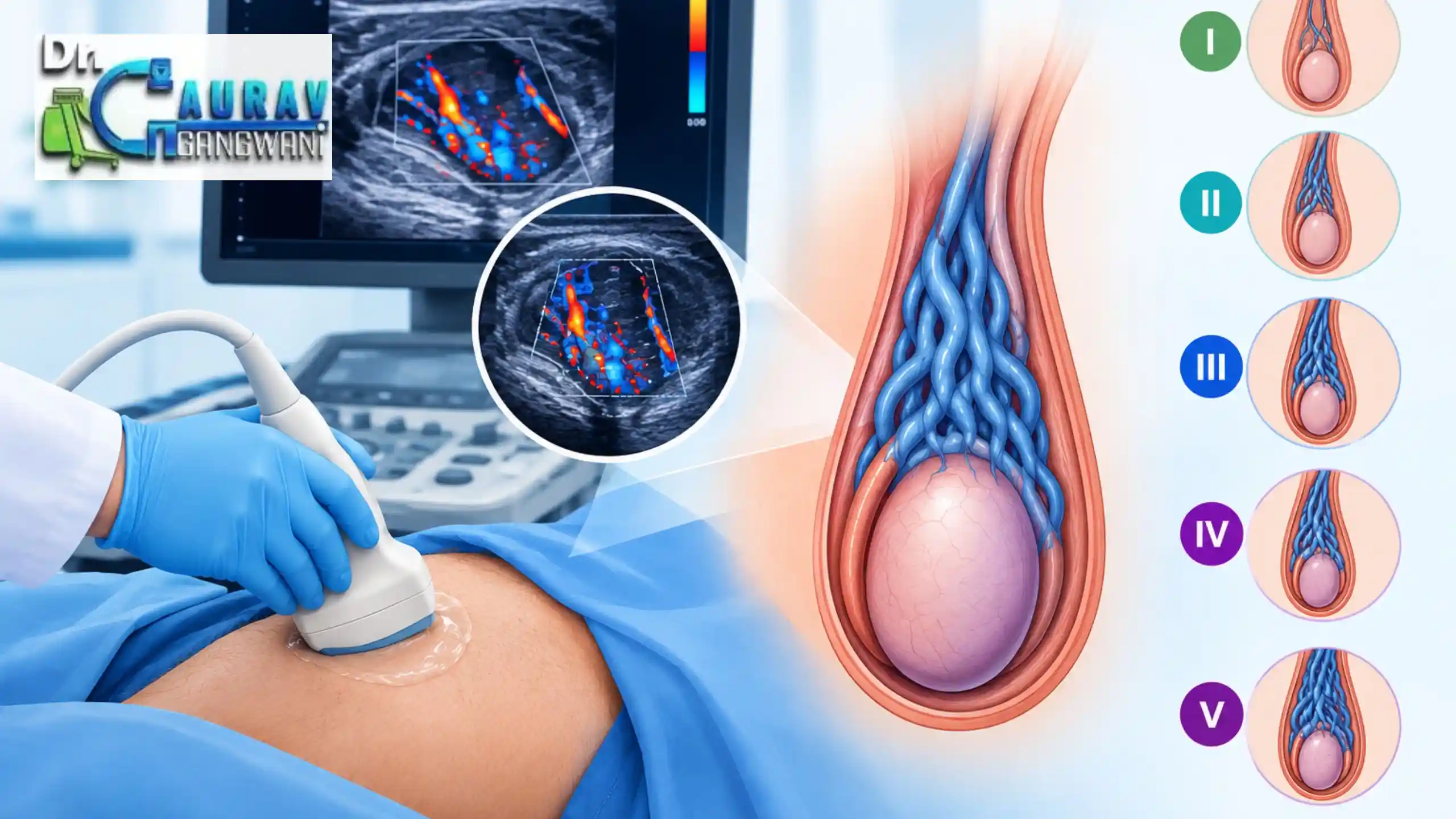
Patients often stare at the words “varicocele grading” on their ultrasound or Doppler report and wonder what it actually means for them. The grade alone doesn’t tell the whole story; it’s just one term doctors use, alongside terms like “enlarged veins” and “blood-flow changes,” to build a full picture of the condition. That said, imaging alone doesn’t decide varicocele treatment it’s considered together with a physical exam, symptoms, fertility concerns, and other clinical findings to determine whether treatment may be appropriate.
The Indian Council of Medical Research includes scrotal Doppler as part of standard evaluation when a varicocele is suspected, so this kind of scan is a normal part of the process, not a cause for alarm.
At Dr. Gaurav Gangwani’s clinic, scan results are explained in plain language and reviewed alongside the patient’s symptoms, helping them understand their report and discuss the right next steps with a specialist.
In radiology, varicocele grading radiology describes the appearance of the affected veins and their blood-flow behavior on ultrasound and color Doppler. Several classification systems have been described, so terminology can vary between reports.
The Sarteschi/Liguori classification is a five-grade ultrasound system that considers the location of venous changes, reflux at rest or during the Valsalva maneuver, and associated testicular findings. There is no universally accepted ultrasound grading system, making the examination technique and reported findings important.
The Sarteschi/Liguori classification breaks varicocele findings down into five grades on ultrasound. What really determines the grade is where the abnormal blood flow (reflux) shows up and whether it’s only triggered by the Valsalva maneuver or already happening while the patient is at rest. As the grade climbs, the findings tend to get more pronounced.
At this earliest stage, reflux only shows up in the veins of the inguinal canal, and only when the patient bears down (Valsalva maneuver). The veins themselves don’t look enlarged yet.
Here, the veins sitting above the testis are noticeably dilated, and reflux appears during Valsalva. The abnormal flow is easier to pick up than at Grade I.
By this point, dilated veins have spread around the testis itself, with reflux still triggered by the Valsalva maneuver.
Resting reflux is present and increases during the Valsalva maneuver. Dilated veins may be seen even at rest, with possible testicular hypotrophy.
Resting reflux is present in the dilated pampiniform plexus and does not increase significantly during Valsalva. Testicular hypotrophy is associated with this grade in the Sarteschi classification.
The grade does not provide the complete ultrasound picture. A standardized Doppler examination also considers the largest vein diameter, location of the veins, patient position, findings at rest and during Valsalva, reflux duration, and testicular volume. Current ultrasound recommendations support assessing both sides in supine and standing positions, using grey-scale, color Doppler, and spectral Doppler where appropriate.
Under the ESUR approach, a vein measuring 3 mm or more in diameter taken while the patient is standing and bearing down (Valsalva) can help support a varicocele diagnosis. But this number shouldn’t be looked at in isolation. It only means something when it’s considered together with the reflux findings and how the exam itself was carried out.
Not necessarily. A higher grade on ultrasound just means the findings look more pronounced; it doesn’t automatically mean treatment is required. Doctors don’t decide on treatment based on the grade alone. They also weigh things like the patient’s symptoms, what shows up on a physical exam, fertility-related concerns, semen analysis results, and several other factors before making that call.
Posture and how the exam is performed can both affect what shows up on Doppler. Because of this, assessment usually involves checking the patient in both supine and standing positions, at rest and during the Valsalva maneuver. Color Doppler demonstrates venous flow, while spectral Doppler can help characterize reflux. ESUR recommendations consider reflux lasting more than two seconds during standing Valsalva abnormal within their standardized approach.
A varicocele grading radiology report is most useful when the grade is read alongside vein diameter, reflux pattern, testicular findings, and examination technique. Because ultrasound classification systems are not completely standardized, the reported grade should not be treated as a standalone measure of severity or fertility impact.
Every grade marks out a distinct pattern of swollen veins and backward blood flow, seen on the varicocele grading radiology Sarteschi/Liguori ultrasound classification.
By Grade 4 and Grade 5, the backward flow shows up even when the patient is resting, so these grades reflect findings that are more constant under this system.
Reflux is just the term for blood moving backward through the veins involved. Where it happens, how long it lasts, and how it changes with a Valsalva maneuver all matter when reading the Doppler results.
Not on its own. Doctors look at the grade together with symptoms, what shows up on physical exam, fertility concerns, and other clinical details before deciding anything.
Ultrasound is good at describing the varicocele itself, but the grade by itself won’t tell you how it’s affecting fertility. For that, a clinical evaluation and the right fertility testing are usually needed too.
Dr. Gangwani Vascular Clinic, 302/TGN SQUARE 3rd floor Chandawarkar lane Borivali west Mumbai 400092
+91 9326195964
drgauravgangwani@gmail.com
Monday to Saturday: 8.30 am to 8.00 pm. For any quires & assistance please call on: +91 9082394787
Copyright 2026 – www.drgauravgangwani.com
Website Managed by SEO Discovery
WhatsApp us