-
Mobile: +91 9082394787
-
Phone: +91 9326195964
-
Email: drgauravgangwani@gmail.com

Osteoid Osteoma Ablation: Radiofrequency Treatment Without Surgery
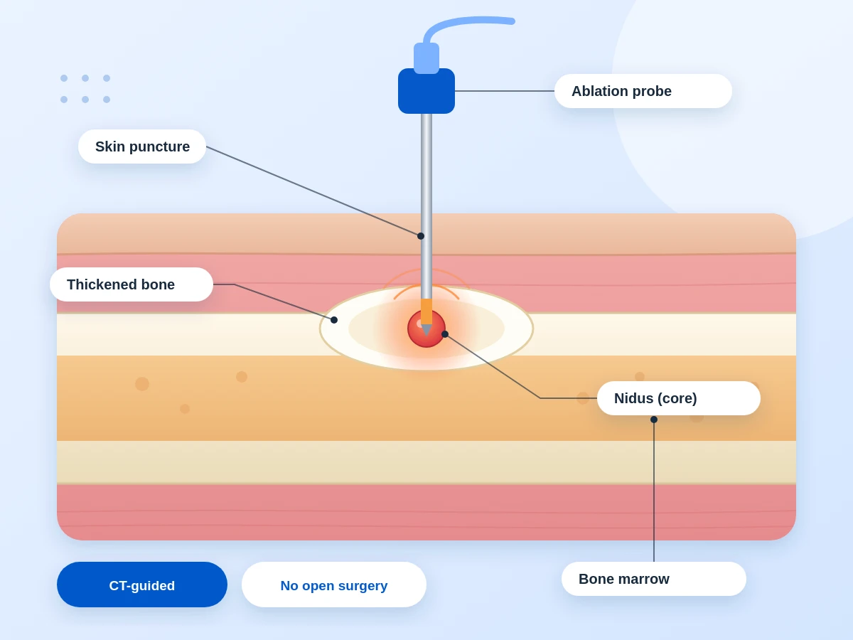
If a deep, nagging pain in one bone keeps disturbing or waking you or your child at night, and painkillers only help for a few hours, a small bone growth called an osteoid osteoma could be the reason. It is not cancer, and it can be treated through a tiny pinhole using heat, with no open surgery. At Dr Gangwani’s Vascular Clinic, we explain osteoid osteoma treatment step by step, so you know exactly what is happening and why.
Understanding the Condition
A Tiny Bone Tumour That Hurts Far More Than Its Size
An osteoid osteoma is a benign (non-cancerous) bone tumour, usually smaller than 2 cm. It doesn’t spread to other bodily areas. It is seen most often in children, teenagers and young adults, and it commonly sits in the thigh bone, shin bone or spine, though any bone can be affected.
At a glance
- Benign: not cancer, and does not spread
- Small: usually under 2 cm
- Who: children, teenagers and young adults
- Where: thigh bone, shin bone or spine
Why does such a small lump hurt so much?
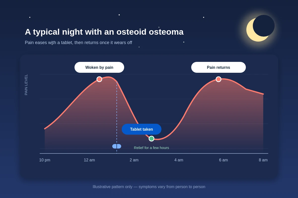
At the centre of the growth is a tiny core called the nidus. It keeps producing inflammatory chemicals that irritate the nerves inside the bone. Anti-inflammatory tablets calm these chemicals for a few hours, which is why the pain eases for a while and then returns. The nidus itself stays active, so the pain keeps coming back until the nidus is treated.
How does an osteoid osteoma show up on a scan?
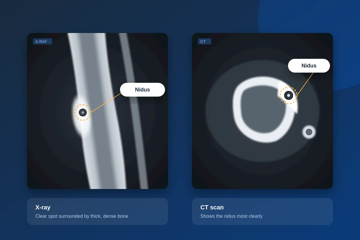
A typical osteoid osteoma X-ray shows a small round or oval clear spot surrounded by thick, dense bone. However, the nidus can be hard to spot, especially in the spine or hip. In that case, your doctor may suggest a CT scan, which shows the nidus most clearly, and sometimes an MRI or bone scan as well.
Warning Signs
Bone Pain in Legs at Night: When Should You See a Specialist?
Most night-time leg aches in children are growing pains or muscle cramps. But if the pain follows a pattern, it is worth getting checked.
Pain that wakes you from sleep
A dull, deep ache in one particular spot, night after night, is a classic warning sign.
Relief with a pain tablet, but only for a while
If an anti-inflammatory such as ibuprofen settles the pain and it returns once the tablet wears off, do not ignore it.
Limping or difficulty with daily life
Trouble sleeping, avoiding sports, limping, or a stiff, painful back in a teenager all need a proper evaluation.
Your Options
Osteoid Osteoma Treatment Options, From Painkillers to Ablation
Any bone pain treatment works well here only when the nidus itself is dealt with. There are three broad routes, and the right one depends on how severe your symptoms are and where the lesion sits.
The older routes: tablets, waiting and open surgery
Long-term painkillers
These control symptoms but do not remove the cause. Using them for months can irritate the stomach.
Waiting it out
Some osteoid osteomas fade on their own, but this can take several years. That is a long time to live with disturbed sleep.
Surgical removal
The surgeon opens the area and removes the nidus. It works, but it means a larger cut, more bone disturbance and a longer recovery.
The Modern Choice
Radiofrequency Ablation for Osteoid Osteoma: The Modern Choice
For persistent pain, image-guided thermal ablation is now an established alternative to surgery. A thin probe is placed into the nidus and heat is used to destroy it. There are two versions: Radiofrequency Ablation (RFA) and Microwave Ablation (MWA). Both work on the same principle. Which one we use depends on the size and location of the lesion, the structures close to it, and the treating specialist’s experience. Neither is better in every case.
Step by Step
How the procedure works, step by step
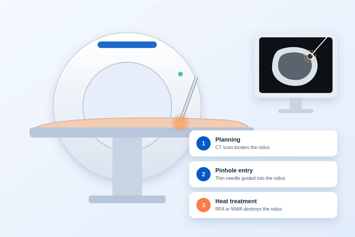
Planning
A CT scan is used to identify the location of the nidus.
Pinhole entry
According to the CT guidance, a thin needle is inserted through a small puncture in the skin into the nidus. Any further special arrangements for anaesthesia for comfort are planned in advance depending on age and location.
Heat treatment
The RFA or MWA probe heats and eliminates the nidus in a controlled manner, with minimal damage to the surrounding bone and tissues.
Key Benefits
Benefits of ablation
No open surgery
Only a pinhole puncture, with no large scar.
Precise treatment
CT guidance means the nidus is targeted without removing healthy bone.
Short stay, quicker recovery
Most people go home soon after the procedure, with excellent pain relief.
After the Procedure
Recovery After Osteoid Osteoma Ablation: What to Expect
Most patients feel a clear improvement in pain soon after a successful procedure and return to normal activities fairly quickly. The exact pace depends on which bone was treated and on your own healing.
The first day or two
The hospital stay is short. You will be advised to rest the treated area initially. If the lesion is in a leg bone, we will tell you how much weight to put on it and when it is safe to walk freely. Mild soreness at the puncture site is common and settles on its own.
Long-term outcomes
- Pain relief: The night pain that disturbed your sleep for months usually settles, and we will guide you on stopping the painkillers.
- Back to routine: School, work and sports are resumed gradually, as advised.
- High success rate: In well-selected patients, osteoid osteoma ablation has high technical and clinical success.
Get the Right Scan First, Then Decide the Next Step
If you or your child has unexplained pain, especially bone pain in the legs at night, the right imaging can tell you whether an osteoid osteoma is the cause. Bring your X-ray, CT or MRI reports along, and our team at Dr Gangwani’s Vascular Clinic will review them and tell you honestly whether ablation is the right osteoid osteoma treatment for your case.
FAQs
FAQs About Osteoid Osteoma Ablation
Is RFA or microwave ablation safe?
Yes, in general. When it is carefully planned and performed under image guidance, thermal ablation is considered a safe, minimally invasive treatment. As with any procedure, your doctor will explain the specific risks for your case.
Does an osteoid osteoma need open surgery?
Not necessarily. For suitable patients, image-guided ablation is a minimally invasive alternative to surgical removal.
Which is better, RFA or MWA?
Both can be effective. The better option depends on where the lesion is, how big it is, how close it is to important structures, and other patient-specific factors.
How long does the procedure take?
It varies with the location and complexity of the lesion. We will give you a realistic estimate once we have seen your scans.
Who is a suitable candidate for osteoid osteoma ablation?
People with a confirmed osteoid osteoma and ongoing symptoms can be considered, after proper imaging and clinical evaluation. Children and adults are both assessed in the same way.
Prime Services
Other Services
Contact Us
Address
Dr. Gangwani Vascular Clinic, 302/TGN SQUARE 3rd floor Chandawarkar lane Borivali west Mumbai 400092
Contact Number
+91 9326195964
Customer Care
drgauravgangwani@gmail.com
Meeting Hours & Video Consultation Timing
Monday to Saturday: 8.30 am to 8.00 pm. For any quires & assistance please call on: +91 9082394787
Copyright 2026 – www.drgauravgangwani.com
Website Managed by SEO Discovery